Marko

Specific inhibition of CK2a from an anchor outside the active site

Paul Brear, Claudia De Fusco, Kathy Hadje Georgiou, Nicola J. Francis-Newton, Christopher J. Stubbs,  Hannah F. Sore, Ashok R. Venkitaraman, Chris Abell, David R. Spring and Marko Hyvönen

Chemical Science, 7(11):6839-6845, 2016
Pubmed: 28451126
DOI:10.1039/C6SC02335E
PDB coordinates:
5CVH (3D view), 5CVG (3D view), 5CVF (3D view), 5CU3 (3D view), 5CU4 (3D view), 5CU6 (3D view), 5CSH (3D view), 5CSP (3D view), 5CSV (3D view), 5CSH (3D view), 5CS6 (3D view), 5CLP (3D view)

Abstract

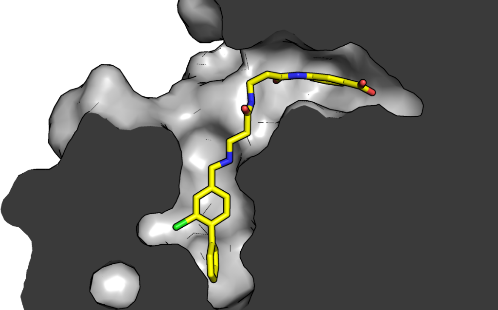
The development of selective inhibitors of protein kinases is challenging because of the significant conservation of the ATP binding site. Here, we describe a new mechanism by which the protein kinase CK2α can be selectively inhibited using features outside the ATP site. We have identified a new binding site for small molecules on CK2α adjacent to the ATP site and behind the αD loop, termed the αD pocket. An elaborated fragment anchored in this site has been linked with a low affinity fragment binding in the ATP site, creating a novel and selective inhibitor (CAM4066) that binds CK2α with a Kd of 320 nM and shows significantly improved selectivity compared to other CK2α inhibitors. CAM4066 shows target engagement in several cell lines and similar potency to clinical trial candidate CX4945. Our data demonstrate that targeting a poorly conserved, cryptic pocket allows inhibition of CK2α via a novel mechanism, enabling the development of a new generation of selective CK2α inhibitors.

Posted by Marko in Publications, 0 comments

Engineering Archeal Surrogate Systems for the Development of Protein-Protein Interaction Inhibitors against Human RAD51

Tommaso Moschetti, Timothy Sharpe, Gerhard Fischer, May E. Marsh, Hong Kin Ng, Matthew Morgan, Duncan E. Scott, Tom L. Blundell, Ashok R. Venkitaraman, John Skidmore, Chris Abell, and Marko Hyvönen

Journal of Molecular Biology 428(23): 4589–4607, 2016

DOI: 10.1016/j.jmb.2016.10.009

PDB coordinates: 5FOS, 5LB2, 5LBI, 5L8V, 5LB4, 5KDD, 5J4L, 5JEE, 5JED, 5JEC, 5JFG, 5J4H, 5J4K


Protein-protein interactions (PPIs) are increasingly important targets for drug discovery. Efficient fragment-based drug discovery approaches to tackle PPIs are often stymied by difficulties in the production of stable, unliganded target proteins. Here, we report an approach that exploits protein engineering to “humanise” thermophilic archeal surrogate proteins as targets for small-molecule inhibitor discovery and to exemplify this approach in the development of inhibitors against the PPI between the recombinase RAD51 and tumour suppressor BRCA2. As human RAD51 has proved impossible to produce in a form that is compatible with the requirements of fragment-based drug discovery, we have developed a surrogate protein system using RadA from Pyrococcus furiosus. Using a monomerised RadA as our starting point, we have adopted two parallel and mutually instructive approaches to mimic the human enzyme: firstly by mutating RadA to increase sequence identity with RAD51 in the BRC repeat binding sites, and secondly by generating a chimeric archaeal human protein. Both approaches generate proteins that interact with a fourth BRC repeat with affinity and stoichiometry comparable to human RAD51. Stepwise humanisation has also allowed us to elucidate the determinants of RAD51 binding to BRC repeats and the contributions of key interacting residues to this interaction. These surrogate proteins have enabled the development of biochemical and biophysical assays in our ongoing fragment-based small-molecule inhibitor programme and they have allowed us to determine hundreds of liganded structures in support of our structure-guided design process, demonstrating the feasibility and advantages of using archeal surrogates to overcome difficulties in handling human proteins.

Pubmed | ScienceDirect

J Mol Biol. 2016 Nov 20;428(23):4589-4607. doi: 10.1016/j.jmb.2016.10.009.

Posted by Marko in Publications, 0 comments

Allosteric modulation of AURKA kinase activity by a small-molecule inhibitor of its protein-protein interaction with TPX2

Matej Janeček, Maxim Rossmann, Pooja Sharma, Amy Emery, David J. Huggins, Simon R. Stockwell, Jamie E. Stokes, Yaw S. Tan, Estrella Guarino Almeida, Bryn Hardwick, Ana J. Narvaez, Marko Hyvönen, David R. Spring, Grahame J. McKenzie & Ashok R. Venkitaraman

Scientific Reports 6, Article number: 28528 (2016)
DOI: 10.1038/srep28528
Pubmed: 27339427

PDB coordinates: 5DNR (3D view ), 5DT3 (3D view ), 5DT0 (3D view ), 5RDR (3D view ), 5DR9 (3D view ), 5DR2 (3D view ), 5DR6 (3D view ), 5DOS (3D view ), 5DPV (3D view ), 5DT4 (3D view ), 5DN3 (3D view )

Abstract

The essential mitotic kinase Aurora A (AURKA) is controlled during cell cycle progression via two distinct mechanisms. Following activation loop autophosphorylation early in mitosis when it localizes to centrosomes, AURKA is allosterically activated on the mitotic spindle via binding to the microtubule-associated protein, TPX2. Here, we report the discovery of AurkinA, a novel chemical inhibitor of the AURKA-TPX2 interaction, which acts via an unexpected structural mechanism to inhibit AURKA activity and mitotic localization. In crystal structures, AurkinA binds to a hydrophobic pocket (the ‘Y pocket’) that normally accommodates a conserved Tyr-Ser-Tyr motif from TPX2, blocking the AURKA-TPX2 interaction. AurkinA binding to the Y- pocket induces structural changes in AURKA that inhibit catalytic activity in vitro and in cells, without affecting ATP binding to the active site, defining a novel mechanism of allosteric inhibition. Consistent with this mechanism, cells exposed to AurkinA mislocalise AURKA from mitotic spindle microtubules. Thus, our findings provide fresh insight into the catalytic mechanism of AURKA, and identify a key structural feature as the target for a new class of dual-mode AURKA inhibitors, with implications for the chemical biology and selective therapeutic targeting of structurally related kinases.

Posted by Marko in Publications, 0 comments

Structure of Gremlin-1 and analysis of its interaction with BMP-2

Miglė Kišonaitė, Xuelu Wang and Marko Hyvönen

Biochemical Journal 473, 1593-1604 (2016)
DOI: 10.1042/BCJ20160254
Pubmed: 27373274
PDB coordinates: 5AEJ (3D view)

Abstract

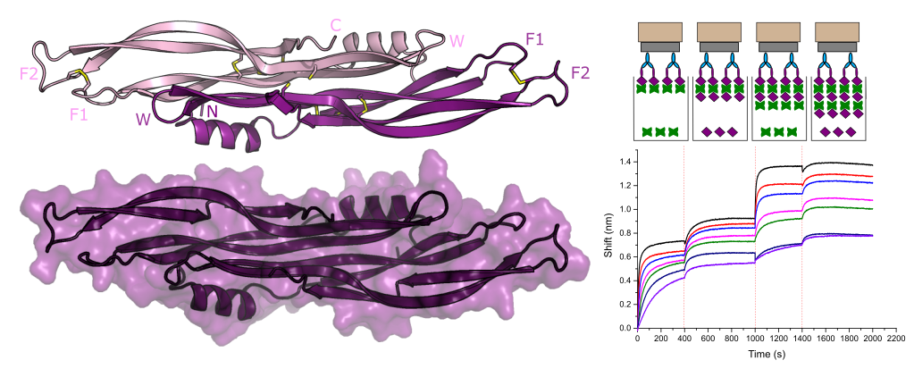
Bone morphogenetic protein 2 (BMP-2) is a member of the transforming growth factor-β (TGF-β) signalling family and has a very broad biological role in development. Its signalling is regulated by many effectors: transmembrane proteins, membrane-attached proteins and soluble secreted antagonists such as Gremlin-1. Very little is known about the molecular mechanism by which Gremlin-1 and other DAN (differential screening-selected gene aberrative in neuroblastoma) family proteins inhibit BMP signalling. We analysed the interaction of Gremlin-1 with BMP-2 using a range of biophysical techniques, and used mutagenesis to map the binding site on BMP-2. We have also determined the crystal structure of Gremlin-1, revealing a similar conserved dimeric structure to that seen in other DAN family inhibitors. Measurements using biolayer interferometry (BLI) indicate that Gremlin-1 and BMP-2 can form larger complexes, beyond the expected 1:1 stoichiometry of dimers, forming oligomers that assemble in alternating fashion. These results suggest that inhibition of BMP-2 by Gremlin-1 occurs by a mechanism that is distinct from other known inhibitors such as Noggin and Chordin and we propose a novel model of BMP-2–Gremlin-1 interaction yet not seen among any BMP antagonists, and cannot rule out that several different oligomeric states could be found, depending on the concentration of the two proteins.

Posted by Marko in Publications, 0 comments

ATP half‐sites in RadA and RAD51 recombinases bind nucleotides

May E. Marsh, Duncan E. Scott, Matthias T. Ehebauer, Chris Abell, Tom L. Blundell and Marko Hyvönen

FEBS OpenBio 6:372–385 (2016)
DOI: 10.1002/2211-5463.12052
Pubmed: 27419043

PDB coordinates: 4UQO (3D view),4D6P (3D view),4B2P (3D view),4A6X (3D view),4A6P (3D view)

Abstract

Homologous recombination is essential for repair of DNA double-strand breaks. Central to this process is a family of recombinases, including archeal RadA and human RAD51, which form nucleoprotein filaments on damaged single-stranded DNA ends and facilitate their ATP-dependent repair. ATP binding and hydrolysis are dependent on the formation of a nucleoprotein filament comprising RadA/RAD51 and single-stranded DNA, with ATP bound between adjacent protomers. We demonstrate that truncated, monomeric Pyrococcus furiosus RadA and monomerised human RAD51 retain the ability to bind ATP and other nucleotides with high affinity. We present crystal structures of both apo and nucleotide-bound forms of monomeric RadA. These structures reveal that while phosphate groups are tightly bound, RadA presents a shallow, poorly defined binding surface for the nitrogenous bases of nucleotides. We suggest that RadA monomers would be constitutively bound to nucleotides in the cell and that the bound nucleotide might play a structural role in filament assembly.

Posted by Marko in Publications, 0 comments

Older Publications

2015

Ultrahigh-throughput discovery of promiscuous enzymes by picodroplet functional metagenomics.

P.-Y. Colin, B. Kintses, F. Gielen, C. M. Miton, G. Fischer, M. F. Mohamed, M. Hyvönen, D. P. Morgavi, D. B. Janssen, F. Hollfelder 
Nature Communications. 6:10008 doi: 10.1038/ncomms10008, 2015.

Alternative Modulation of Protein-Protein Interactions by Small Molecules.

G. Fischer, M. Rossmann, M. Hyvönen
Current Opinion in Biotechnology, 35:78-85 doi: 10.1016/j.copbio.2015.04.006, 2015.

2014

Small molecule inhibitors targeting protein-protein interaction in the RAD51 family of recombinases

D.E. Scott, A. G. Coyne, T. L. Blundell, A. Venkitaraman, C. Abell, M. Hyvönen
ChemMedChem, 10: 296–303 DOI: 10.1002/cmdc.201402428, 2014

Functionalised staple linkages for modulating the cellular activity of stapled peptides.

Y. H. Lau, P. de Andrade, S.-T. Quah, M. Rossmann, L. Laraia, N. Sköld, T. J. Sum, P. J. E. Rowling, T. L. Joseph, C. Verma, M.Hyvönen, L. S. Itzhaki, A. R. Venkitaraman, C. J. Brown, D. P. Lane, D. R. Spring
Chemical Science, 5:18040-1809, doi: 0.1039/C4SC00045E, 2014

2012

Targeting the RAD51:BRCA2 Protein-Protein Interaction using Fragment-based methods.

D. E. Scott, M. T. Ehebauer, T. Pukala, M. Marsh, T. L. Blundell, A. R. Venkitaraman, C. Abell, M. Hyvönen. ChemBioChem,14:332-42, 2013
A. Sharma, F. Meyer, M.Hyvönen, S. M. Best, R. E. Cameron, N. Rushton. Osteoinduction by combining bone morphogenetic protein (BMP)-2 with a bioactive novel nanocomposite
Bone and Joint Research. 1:145-51, 2012

2011

Targeting protein-protein interactions and fragment-based drug discovery.

E. Valkov, T. Sharpe, M. Marsh, S. Greive and M. Hyvönen
In Topics in Current Chemistry: “Fragment-based drig discovery and X-ray crystallography” ed. T. Davies and M. Hyvönen, 2011, DOI: 10.1007/128_2011_265

From crystal packing to molecular recognition: prediction and discovery of a binding site on the surface of polo-like kinase 1.

P Sledź, C. J. Stubbs, S. Lang, Y. Q. Yang, G. J. McKenzie, A. R. Venkitaraman, M. Hyvönen, C Abell
Angewante Chemie International Edition, 50:4003-6, 2011

2010

An efficient,multiply promiscuous hydrolase in the alkaline phosphatase superfamily.

B. van Loo, S. Jonas, A. C. Babtie, A. Benjdia, O. Berteau, M. Hyvönen, and F. Hollfelder
Proceedings of the National Academy of Sciences of the USA, 107:2740-5, 2010

2009

Rab5-mediated endocytosis of activin is not required for gene activation or long-range signalling in Xenopus.

A. I. Hagemann, X. Xu, O. Nentwich, M. Hyvönen, K. Dingwell and J. C. Smith
Development, 136:2803-13, 2009

A new ‘total’ activin B ELISA: development and validation for human samples.

H. Ludlow, D. J. Phillips, M. Myers, R. I. McLachlan, D. M. de Kretser, C. A. Allan, R. A. Anderson, N. P. Groome, M. Hyvönen, W. C. Duncan and S. Muttukrishna
Clinical Endocrinology. 71:867-73, 2009

2008

A new member of the alkaline phosphatase superfamily with a formylglycine nucleophile: structural and kinetic characterisation of a phosphonate monoester hydrolase/phosphodiesterase from Rhizobium leguminosarum.

S. Jonas, B. van Loo, M. Hyvönen, F. Hollfelder
Journal of Molecular Biology, 384:120-36, 2008

Development of a new antibody to the human inhibin/activin βB subunit and its application to improved inhibin B ELISAs.

H. Ludlow, S. Muttukrishna, M. Hyvönen , N.P. Groome
Journal of Immunological Methods, 329:102-111, 2008

2006

Structural basis for the inhibition of activin signalling by follistatin.

A.E. Harrington, S.A. Morris-Triggs, B.T. Ruotolo, C.V. Robinson, S. Ohnuma and M. Hyvönen
EMBO Journal, 25:1035-1045, 2006

2003

CHRD, a novel domain in the bone morphogenetic protein inhibitor chordin, is also found in microbial proteins.

M. Hyvönen
Trends in Biochemical Sciences, 28: 470–473, 2003

Adhesion of endothelial cells to nov is mediated by integrins αv3 and β51.

P. D. Ellis, J. C. Metcalf, M. Hyvönen, and P. R. Kemp
Journal of Vascular Research, 40:234–243, 2003

Crystal structures of the heparan sulfate-binding domain of follistatin. Insights into ligand binding.

C. A. Innis and M. Hyvönen
Journal of Biological Chemistry, 278:39969–39977, 2003

2000

Protein-protein interactions in eukaryotic signal transduction.

M. Hyvönen, J. Begun, and T. Blundell
In Protein-Protein Recognition, Frontiers in Molecular Biology, pages 189–227. Oxford University Press, 2000

1999

Structure of the PH domain from Bruton’s tyrosine kinase in complex with inositol 1,3,4,5-tetrakisphosphate.

E. Baraldi, K. Djinovic-Carugo, M. Hyvönen, P. Lo Surdo, A. M. Riley, B. V. L. Potter, R. O’Brien, J. E. Ladbury, and M. Saraste
Structure, 7:449–460, 1999

Expression of cDNAs in Escherichia coli using T7 promoter.

M. Hyvönen and M. Saraste.
In Cell Biology – A laboratory manual, volume 4, pages 255–261. Academic Press, 2nd edition, 1997a

Structure of PH domain and Btk motif from Bruton’s tyrosine kinase: molecular explanations for the X-linked agammaglobulinemia.

M. Hyvönen and M. Saraste
EMBO Journal, 16:3396–3404, 1997

Structure of the WW domain of a kinase-associated protein complexed with a proline-rich peptide.

J. Macias, M. Hyvönen, E. Baraldi, J. Schultz, M. Sudol, M. Saraste, and H. OschkinatNature, 382:646–649, 1996.

T7 vectors with modified T7lac promoter for expression of proteins in Escherichia coli.

J. Peränen, M. Rikkonen, M. Hyvönen, and L. Kääriäinen
Analytical Biochemistry, 236:371–373, 1996.

The alphavirus replicase protein nsP1 is membrane-associated and has affinity to endocytic organelles.

J. Peränen, P. Laakkonen, M. Hyvönen, and L. Kääriäinen
Virology, 208:610–620, 1995.

Structure of the binding site for inositol phosphates in a PH domain.

M. Hyvönen, M. J. Macias, M. Nilges, H. Oschkinat, M. Saraste, and M. Wilmanns
EMBO Journal, 14:4676–4685, 1995.

Pleckstrin homology domains: a fact file.

M. Saraste and M. Hyvönen
Current Opinion in Structural Bioloy, 5:403–408, 1995.

The C-terminal domain of α-spectrin is structurally related to calmodulin.

G. Trave, A. Pastore, M. Hyvönen, and M. Saraste
European Journal of Biochemistry, 227:35–42, 1995.

PH domain: the first anniversary.

T.J. Gibson, M. Hyvönen, A. Musacchio, M. Saraste, and E. Birney
Trends in Biochemical Sciences, 19:349–353, 1994.

Expression of Semliki forest virus nsP1-specific methyltransferase in insect cells and in Escherichia coli.

P. Laakkonen, M. Hyvönen, J. Peränen, and L. Kääriäinen
Journal of Virology, 68:7418–7425, 1994.
Posted by Marko in Publications, 0 comments