Marko

Second-generation CK2α inhibitors targeting the αD pocket

Jessica Iegre, Paul Brear, Claudia De Fusco, Masao Yoshida, Sophie L. Mitchell, Maxim Rossmann, Laura Carro Santos, Hannah F. Sore, Marko Hyvӧnen and David R. Spring

Chemical Science (2018) (9:3041-)
DOI: 10.1039/C7SC05122K
Pubmed: 29732088

PDB coordinates:

6EHU (3D view ), 5OTQ (3D view ), 5OTY (3D view ), 6EHK (3D view ), 5OTI (3D view ), 5OTL (3D view ), 5OTO (3D view ), 5OYF (3D view ), 5OSZ (3D view ), 5OT5 (3D view ), 5OTD (3D view ), 5OTH (3D view ), 5OT6 (3D view ), 5OUE (3D view ), 5OUM (3D view ), 5OUU (3D view ), 5OS8 (3D view ), 5OTR (3D view ), 6EII (3D view ), 5OQU (3D view ), 5ORK (3D view ), 5OSL (3D view ), 5OUL (3D view ), 5ORH (3D view ), 5ORJ (3D view ), 5OS7 (3D view )

Abstract

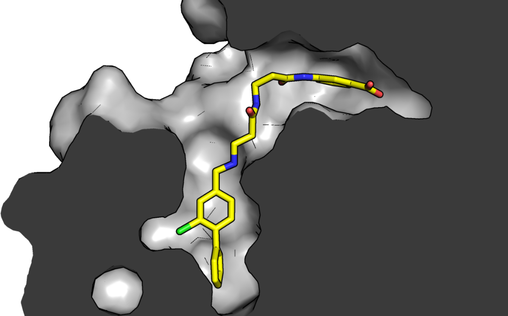
CK2 is a critical cell cycle regulator that also promotes various anti-apoptotic mechanisms. Development of ATP-non-competitive inhibitors of CK2 is a very attractive strategy considering that the ATP binding site is highly conserved among other kinases. We have previously utilised a pocket outside the active site to develop a novel CK2 inhibitor, CAM4066. Whilst CAM4066 bound to this new pocket it was also interacting with the ATP site: herein, we describe an example of a CK2α inhibitor that binds completely outside the active site. Continue reading →

Posted by Marko in Publications, 0 comments

Structural and Mechanistic Analysis of the Choline Sulfatase from Sinorhizobium melliloti: A Class I Sulfatase Specific for an Alkyl Sulfate Ester

Bert van Loo, Markus Schober, Eugene Valkov, Magdalena Heberlein, Erich Bornberg-Bauer, Kurt Faber, Marko Hyvönen, Florian Hollfelder

Journal of Molecular Biology, 430:1004-1023, 2018 
DOI: 10.1016/j.jmb.2018.02.010
Pubmed: 29458126
PDB coordinates:
6FNY (3D view )


Abstract

Hydrolysis of organic sulfate esters proceeds by two distinct mechanisms, water attacking at either sulfur (S-O bond cleavage) or carbon (C-O bond cleavage). In primary and secondary alkyl sulfates attack at carbon is favored, whereas in aromatic sulfates and sulfated sugars attack at sulfur is preferred. This mechanistic distinction is mirrored in the classification of enzymes that catalyze sulfate ester hydrolysis: arylsulfatases catalyze S-O cleavage in sulfate sugars and arylsulfates and alkyl sulfatases break the C-O bond of alkyl sulfates. Continue reading →

Posted by Marko in Publications, 0 comments

Molecular characterization of latent GDF8 reveals mechanisms of activation

Ryan G. Walker, Jason C. McCoy, Magdalena Czepnik, Melanie J. Mills, Adam Haggd, Kelly L. Walton, Thomas R. Cotton, Marko Hyvönen, Richard T. Lee, Paul Gregorevic, Craig A. Harrison, and Thomas B. Thompson


Pubmed:29348202

Abstract

Growth/differentiation factor 8 (GDF8), or myostatin, negatively regulates muscle mass. GDF8 is held in a latent state through interactions with its N-terminal prodomain, much like TGF-β. Using a combination of small-angle X-ray scattering and mutagenesis, we characterized the interactions of GDF8 with its prodomain. Our results show that the prodomain:GDF8 complex can exist in a fully latent state and an activated or “triggered” state where the prodomain remains in complex with the mature domain. Continue reading →

Posted by Marko in Publications, 0 comments

Structure of the human pro-myostatin precursor and determinants of growth factor latency

Thomas Cotton, Gerhard Fischer, Xuelu Wang, Jason McCoy, Magdalena Czepnik, Thomas B. Thompson, Marko Hyvönen

The EMBO Journal (2018) 37: 367-383
DOI: 10.15252/embj.201797883
Pre-publication in BiorXiv, DOI: doi.org/10.1101/153403
PDB coordinates: 5NTU (3D view ), 5NXS (3D view )


Abstract

Myostatin, a key regulator of muscle mass in vertebrates, is biosynthesised as a latent precursor in muscle and is activated by sequential proteolysis of the pro-domain. To investigate the molecular mechanism by which pro-myostatin remains latent, we have solved the structure of unprocessed pro-myostatin and analysed the properties of the protein in its different forms. Continue reading →

Posted by Marko in Publications, 0 comments
Paul & co’s work on CK2α inhibition highlighted

Paul & co’s work on CK2α inhibition highlighted

Dan Erlanson has highlighted our project on Ck2α inhibitor development in his Practical Fragments blog  “Fragment Linking to selective Ck2 inhibitor“.

This a project we have done in close collaboration with David Spring’s group at the Department of Chemistry. The story started from a serendipitous observation that a fragment expected to bind on the top of the N-lobe of CK2α, on the interaction site with scaffolding protein Ck2β bound in a number of different sites on the kinase, including in a new pocket close to the ATP binding site of the kinase. This previously unidentified site was observed only thanks to a new crystal form Paul had obtained. In this crystal form, the so-called αD helix was mobile enough to be displaced by a fragment that, soaked at high concentration,  found a new home in the very hydrophobic pocket that the displacement of the helix revealed. After some optimisation of the fragment and a crystallographic screen to identify ATP-site binding “war heads”, Claudia and others in the Spring lab were able to create the linked molecule CAM4066.

The main story was published in Chemical Science and the detailed description of the design process came out this year in Bioorganic and Medicinal Chemistry.

If interested more in this story, do check in Youtube some of the movies Paul has made from this project:

Development of CAM4066

Optimisation of the fragment in the aD pocket

Growth of the linker from the aD site to the active site

And it should not go forgotten that all the work has been guided continuous crystallographic assessement of the process, with ~30 unique crystal structures in the two papers: 5CVH (3D view), 5CVG (3D view), 5CVF (3D view), 5CU3 (3D view), 5CU4 (3D view), 5CU6 (3D view), 5CSH (3D view), 5CSP (3D view), 5CSV (3D view), 5CSH (3D view), 5CS6 (3D view), 5CLP (3D view), 5MMF (3D view ), 5MMR (3D view ), 5MO5 (3D view ), 5MO6 (3D view ), 5MO7 (3D view ), 5MO8 (3D view ), 5MOD (3D view ), 5MOE (3D view ), 5MOH (3D view ), 5MOT (3D view ), 5MOV (3D view ), 5MOW (3D view ), 5CU0 (3D view ), 5CU2 (3D view ), 5CT0 (3D view ), 5CTP (3D view ), 5CX9 (3D view ).

And there is more to come! Keep your eyes open.

Posted by Marko in News

Computationally-guided optimization of small-molecule inhibitors of the Aurora A kinase–TPX2 protein–protein interaction

Daniel J. Cole, Matej Janecek, Jamie E. Stokes, Maxim Rossmann, John C. Faver, Grahame J. McKenzie, Ashok R. Venkitaraman, Marko Hyvönen, David R. Spring, David J. Huggins and William L. Jorgensen

Chemical Communications 53, 9372-9375 (2017)
DOI 10.1039/C7CC05379G
Pubmed: 28787041

PDB coordinates: 5OBR (3D view)

Abstract

Free energy perturbation theory, in combination with enhanced sampling of protein–ligand binding modes, is evaluated in the context of fragment-based drug design, and used to design two new small-molecule inhibitors of the Aurora A kinase–TPX2 protein–protein interaction.

Posted by Marko in Publications, 0 comments

A fragment-based approach leading to the discovery of a novel binding site and the selective CK2 inhibitor CAM4066.

Claudia De Fusco, Paul Brear, Jessie Iegre, Kathy H. Georgiou, Hannah F. Sore, Marko Hyvönen, David R. Spring

Bioorganic and Medicinal Chemistry, 25:3471-3482 (2017)
DOI: 10.1016/j.bmc.2017.04.037
Pubmed: 28495381

PDB coordinates: 5MMF (3D view ), 5MMR (3D view ), 5MO5 (3D view ), 5MO6 (3D view ), 5MO7 (3D view ), 5MO8 (3D view ), 5MOD (3D view ), 5MOE (3D view ), 5MOH (3D view ), 5MOT (3D view ), 5MOV (3D view ), 5MOW (3D view ), 5CU0 (3D view ), 5CU2 (3D view ), 5CT0 (3D view ), 5CTP (3D view ), 5CX9 (3D view )

Abstract

Recently we reported the discovery of a potent and selective CK2α inhibitor CAM4066. This compound inhibits CK2 activity by exploiting a pocket located outside the ATP binding site (αD pocket). Here we describe in detail the journey that led to the discovery of CAM4066 using the challenging fragment linking strategy. Specifically, we aimed to develop inhibitors by linking a high-affinity fragment anchored in the αD site to a weakly binding warhead fragment occupying the ATP site. Moreover, we describe the remarkable impact that molecular modelling had on the development of this novel chemical tool. The work described herein shows potential for the development of a novel class of CK2 inhibitors.

Posted by Marko in Publications, 0 comments

Structural analyses of von Willebrand factor C domains of collagen 2A and CCN3 reveal an alternative mode of binding to bone morphogenetic protein-2

Emma-Ruoqi Xu, Emily E. Blythe, Gerhard Fischer and  Marko Hyvönen

Journal of Biological Chemistry, 292:12516-12527, 2017
doi:10.1074/jbc.M117.788992
Pubmed ID: 28584056
PDB coordinates: 5NIR (3D view), 5NB8 (3D view)

Abstract:

Bone morphogenetic proteins (BMPs) are secreted growth factors that promote differentiation processes in embryogenesis and tissue development. Regulation of BMP signalling involves binding to a variety of extracellular proteins, among which are many von Willebrand factor C (vWC) domain-containing proteins. While the crystal structure of the complex of crossveinless-2 (CV-2) vWC1 and BMP-2 previously revealed one mode of the vWC:BMP binding mechanism, other vWC domains may bind to BMP differently.  Continue reading →

Posted by Marko in Publications, 0 comments

Development of a multipurpose scaffold for the display of peptide loops

Maxim Rossmann, Sandra J. Greive, Tommaso Moschetti, Michael Dinan, Marko Hyvönen

Protein Engineering, Design and Selection 30: 419–430 (2017)
DOI: 10.1093/protein/gzx017
Pubmed: 228444399

Abstract

Protein-protein interactions (PPIs) determine a wide range of biological processes and analysis of these dynamic networks is increasingly becoming a mandatory tool for studying protein function. Using the globular ATPase domain of recombinase RadA as a scaffold, we have developed a peptide display system (RAD display), which allows for the presentation of target peptides, protein domains or full-length proteins and their rapid recombinant production in bacteria. Continue reading →

Posted by Marko in Publications, 0 comments

Structure and activation of pro-activin A

Xuelu Wang, Gerhard Fischer and Marko Hyvönen

Nature Communications 7:1205, 2016
DOI: 10.1038/ncomms12052
Pubmed: 27373274

PDB coordinates: 5HLY (3D view), 5HLZ (3D view)

Abstract

Activins are growth factors with multiple roles in the development and homeostasis. Like all TGF-β family of growth factors, activins are synthesized as large precursors from which mature dimeric growth factors are released proteolytically. Here we have studied the activation of activin A and determined crystal structures of the unprocessed precursor and of the cleaved pro-mature complex. Replacing the natural furin cleavage site with a HRV 3C protease site, we show how the protein gains its bioactivity after proteolysis and is as active as the isolated mature domain. The complex remains associated in conditions used for biochemical analysis with a dissociation constant of 5 nM, but the pro-domain can be actively displaced from the complex by follistatin. Our high-resolution structures of pro-activin A share features seen in the pro-TGF-β1 and pro-BMP-9 structures, but reveal a new oligomeric arrangement, with a domain-swapped, cross-armed conformation for the protomers in the dimeric protein.

Posted by Marko in Publications, 0 comments